01
Jan
X-Rays

When X-rays pass through your mouth during a dental exam, more X-rays are absorbed by the denser parts (such as teeth and bone) than by soft tissues (such as cheeks and gums) before striking the film. This creates an image on the radiograph. Teeth appear lighter because fewer X-rays penetrate to reach the film. Cavities and gum disease appear darker because of more X-ray penetration. The interpretation of these X-rays allows the dentist to safely and accurately detect hidden abnormalities.
How often dental X-rays (radiographs) should be taken depends on the patient`s individual health needs. It is important to recognize that just as each patient is different from the next, so should the scheduling of X-ray exams be individualized for each patient. Your medical and dental history will be reviewed and your mouth examined before a decision is made to take X-rays of your teeth.
Request an Appointment
The schedule for needing radiographs at recall visits varies according to your age, risk for disease and signs and symptoms. Recent films may be needed to detect new cavities, or to determine the status of gum disease or for evaluation of growth and development. Children may need X-rays more often than adults. This is because their teeth and jaws are still developing and because their teeth are more likely to be affected by tooth decay than those of adults.
Dental X-ray FAQs
During your routine exam, your dentist may request X-rays as part of your check-up. Many patients usually have questions about dental X-rays, and we’re sure you do as well. You have questions, and we have answers. Keep reading the FAQs section below to learn more about dental X-rays.
Dental X-rays, or dental radiographs, use electromagnetic radiation to take images of the mouth, including your teeth, gums, and jawbones. Dense structures like teeth and bones absorb more X-rays, appearing as lighter areas on the X-ray image. In contrast, soft tissues, like gums, appear darker in the image because they allow more X-rays to pass through.
A dental exam comprises two parts. The visual and physical exam inspects the visible areas of the mouth for signs of decay, misalignment, gum disease, etc. The second part of the dental exam is dental X-rays, which are used to examine your dental health beneath the gum surface. Dental X-rays can help unearth numerous dental issues, including bone loss, impacted teeth, cysts, hidden cavities, etc. We don’t always use dental X-rays when we suspect something is wrong. X-rays are preventive, providing early warnings of dental issues before they grow bigger.
Several types of dental X-rays exist. Bitewing X-rays focus on the upper and lower molars and premolars, showing the crowns of the teeth and the supporting bone. These X-rays are used to spot dental decay. Periapical X-rays capture the entire tooth—from root to crown—and the adjacent bone. These X-rays are ideal for detecting tooth infections.
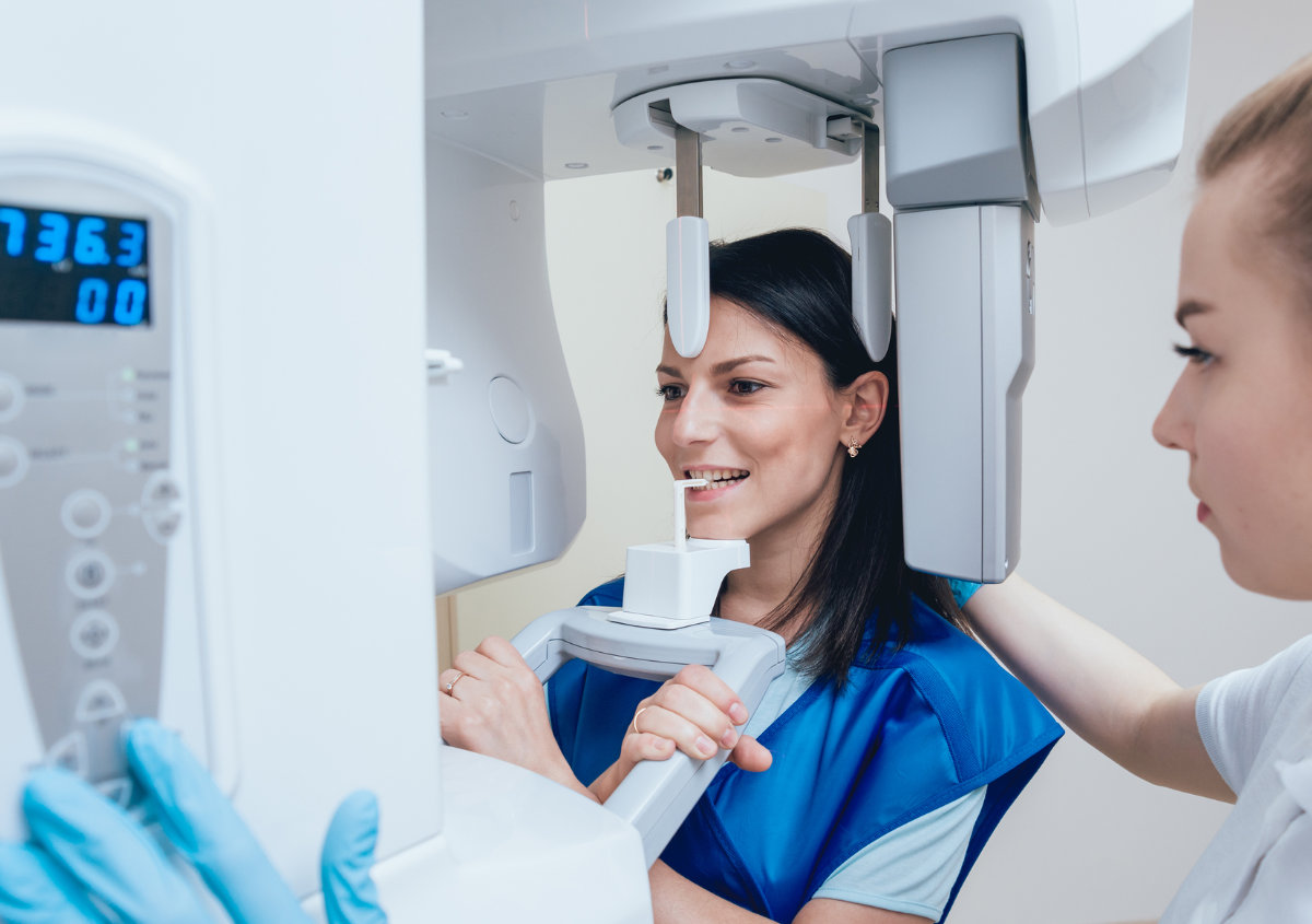
Panoramic X-rays reveal a broader view of the mouth, including all teeth, jaws, and surrounding structures. These X-rays provide advanced diagnostics and are vital for dental implant planning and diagnosing TMJ pain. Lastly, a Cone Beam 3D CT scan allows the dentist to view your oral structures in 3D, allowing precise dental implant surgery and spotting issues missed by conventional X-rays.
Whatever the X-ray type, the process is painless and fast. No noisy machinery is involved, and the process takes 10 minutes or less. Furthermore, there is little preparation required. We’ll give you protective gear to ensure only your mouth is exposed to the X-rays.
Douglas Hoppe DDS uses digital X-rays or radiographs, which differs from traditional films used years ago. Digital radiography offers many benefits, including:
• Faster processing time
• High-quality images
• Low radiation levels
• Eco-friendly
• Easy transfer and storage of medical records
The frequency of dental X-rays depends on the patient. Some patients only need X-rays once annually or every two years. Others need them every six months or more frequently, depending on the developing issues.
We won’t suggest dental X-rays for the sake of it. We’ll evaluate the benefits and risks involved before recommending dental X-rays. Rest assured, we’ll recommend X-rays for a good reason.
Radiation in X-rays is usually one of the main concerns of many patients. The truth is that, unlike film-based dental X-rays, modern radiographs emit minimal radiation levels. Plus, we’ll observe the necessary precautions, including wearing protective gear, to ensure minimal exposure to other body parts. Furthermore, you’ll only get X-rays when needed to avoid unnecessary risk.
If you have further questions about dental X-rays, call (517) 667-7066 to talk to Dr. Douglas Hoppe and the team in Eaton Rapids, MI. We are proud to create healthy, beautiful smiles with innovative technology.
Share this Article
What Our
Patients Say
We always want to assure that our patients receive great care and have good experience when they come to see us.


